
 Harvard Medical School researchers Bock et al took a mouse - just one - and used two forms of microscopy to investigate a small patch of it's primary visual cortex, the area which receives input from the eyes.
First, they used two-photon calcium imaging to look at the functional properties of individual cells. They displayed various kinds of patterns in front of the mouse's eyes, and looked to see which cells lit up, using a special dye which become fluorescent in the presence of calcium, which rises inside cells when they fire.
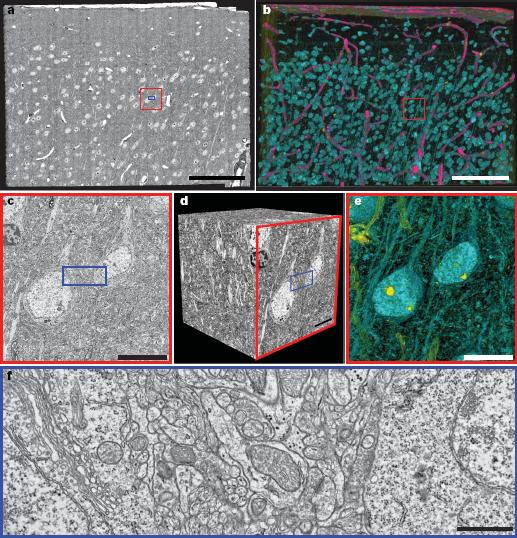
Having done that they took the same chunk of cortex (a rough cube of about 0.4 mm on each side) and used electron microscopy to see it in its entirety. This was the tricky part. Electron microscopy only works if the sample is first cut into extremely thin slices. Each of the 1,200 slices took 20 minutes to image so in total they spent "several months" to get it all done, using a home-made device consisting of 4 high-resolution digital cameras that fed the information to an image processing system.
In total, they acquired 36 terabytes of electron microscope images, and after processing it all they ended up with a 3D picture of 10 million megapixels. My phone has a 16 GB internal storage and a 5 megapixel camera, so in order to get this data I would have to take 2 million photos, and it would take over 2000 phones to store them. There isn't an app for that...yet.
The end result was some very pretty pictures, and amazing movies. Oh, and also, some science - they were able to compare the functional properties of brain cells to their actual physical wiring diagram. This, in the broadest sense, is what all neuroscientists are trying to do; Bock et al, however, went out and did it directly.
 They were able to test an important hypothesis, namely that in the visual cortex, pyramidal cells (the main cortical cell type) project to inhibitory GABA interneurons in a manner which doesn't depend on their orientation-selectivity - whether they respond most strongly to seeing vertical lines, horizontal lines, diagonal ones, etc. Bock et al found that this seemed to be true: pyramidal cells synapsed onto whichever GABA cells happened to be nearest to them, regardless of their orientation-selectivity.
Still, it took them several months to image an area containing just 1,000 neurons. The mouse cortex has 4 million, and the human cortex has 11,000 million, so this is a tiny fraction of the whole brain, and the small size of the area meant that they were only able to examine short-range connections between neighbouring cells, not long-range wiring. So this is early days, but it's clearly an extremely exciting technique and is sure to open the way to major advances in the future.
Link: Also blogged at Brains Lab.
 Bock DD, Lee WC, Kerlin AM, Andermann ML, Hood G, Wetzel AW, Yurgenson S, Soucy ER, Kim HS, & Reid RC (2011). Network anatomy and in vivo physiology of visual cortical neurons. Nature, 471 (7337), 177-82 PMID: 21390124
Tidak ada komentar:
Posting Komentar